Embryonic development of cartilaginous fish

Small-spotted catshark. [a]
This page outlines the embryonic development of the major group of cartilaginous fish known as chondrichthyans, the main subgroup of which is the elasmobranches which comprises sharks and rays. The following description is based primarily on the small-spotted catshark (Scyliorhinus canicula, also known as the lesser-spotted dogfish) which is thought to be fairly typical of elasmobranch embryonic development.
Egg and fertilisation
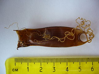
Catshark egg, sometimes known as a ‘Mermaid’s Purse’. Note the very long tendrils which can be up to 1 m. [b]
Elasmobranch eggs are generally quite large and contained within a collagen-based case. The female pronucleus is inside a germinal disc which is orange and located on the outer surface of the yolk, the bulk of which is typically pale yellow-green, except for immediately around the germinal disc where it is white.
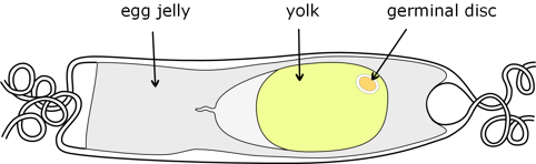
Figure 1. Schematic of catshark egg.
The egg is fertilised while it is within the female oviduct, before addition of the outer case. In elasmobranches it is common for many (up to 50) sperm to enter the germinal disc, although only one combines with the female pronucleus to form the zygote. The early stages of embryonic development take place while the egg is within the oviduct, with the egg reaching about the blastula stage by the time it is released.
Cleavage and blastula
Typical of eggs having a large yolk, cleavage is meroblastic, which means that the early cell divisions do not penetrate the yolk, but are confined to the germinal disc. So in elasmobranches cleavage results in the germinal disc (now called a blastodisc) becoming a clump of cells (called blastomeres) lying within a depression on the surface of the yolk, rather than the egg as a whole becoming a ball of cells.
Early cell divisions result in progressively smaller cells, rather than an increase in size of the blastodisc which remains surrounded by a layer of white yolk. Also in elasmobranches, in the early stages the formation of cell walls lags behind divisions of the cell nuclei, resulting in many new cell nuclei before there are any discrete cells. Even after complete cells have begun to form, a substantial part of the blastodisc, especially on the lower or yolk side is cytoplasm with many nuclei – called a yolk syncytial layer (YSL). (Because of polyspermy, in the early stages there are also many male pronuclei, but these gradually degrade and do not contribute to the embryo.)
Up to about 100 in number the cells are loosely aggregated, but they gradually compact to leave a subgerminal cavity beneath the cells. The surface layer of cells becomes a flattened epithelium, whereas those below are mesenchymal. It is now called a blastula.

Figure 2. Section through early catshark blastula.
Initially the subgerminal cavity is located at the posterior end of the embryo, but then both the surface layer of cells and those beneath it spread to cover the whole of the white yolk.
For a fate map of the blastula see here.
Gastrulation
Towards the rear of the blastodisc, the surface layer of cells first becomes thicker, and then folds under to form a double layer which overhangs the underlying yolk (Figure 3). The upper layer is sometimes called the epiblast, and the lower layer referred to as hypoblast (but note these are different from the epiblast and hypoblast of amniotes which form before gastrulation).

Figure 3. Section through embryonic overhang.
Viewed from above this posterior overhang has two arms, one either side of what is called the ‘notochordal triangle’.
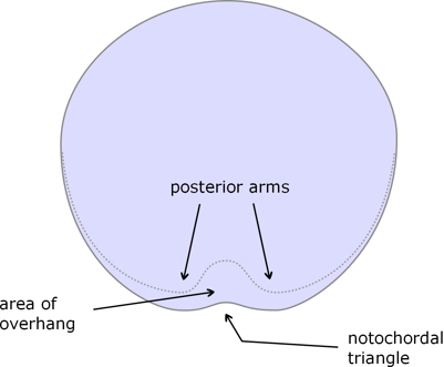
Figure 4. Catshark blastula seen from above, showing the beginning of the embryonic shield.
This overhang, often called the embryonic shield, then extends posteriorly: partly by continued spreading of the upper layer, with further of its cells rolling over the posterior edge, and partly through proliferation of the cells in the lower layer.
At first the cells of both upper and lower layers of the overhang are epithelial in nature, but they progressively thicken and differentiate:
- the upper layer into ectoderm, and
- the lower layer into mesoderm and endoderm.
The ectoderm near the midline first thickens and then becomes narrower laterally but longer (called convergent extension) to form the neural plate, the forerunner of the neural tube (see below), whereas the mesoderm along the embryo’s midline becomes the notchord. Figure 5 is a longitudinal section through the midline after the early stages of gastrulation to indicate the destinies of the cell layers.
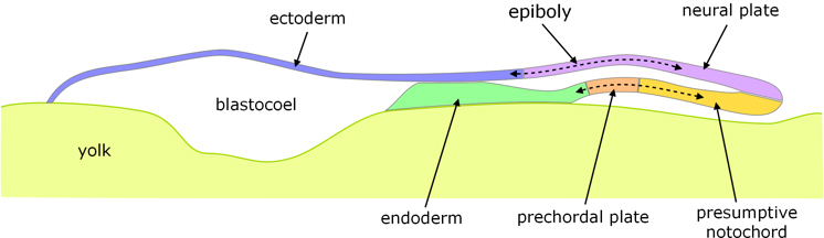
Figure 5. Longitudinal section of gastrulation in the catshark.
As well as differentiating along the anterior-posterior axis, the layers also differentiate laterally, as indicated in figure 6.
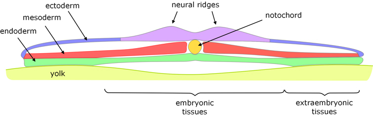
Figure 6. Transverse section following gastrulation, taken though the body of the gastrula rather than the overhang to show lateral differentiation and the approximate demarcation between embryonic and extraembryonic tissues (see 'yolk membranes' below).
Neural tube
The edges of the neural plate thicken to form neural ridges (with the neural groove in-between) which rise on either side and meet to form the neural tube. On meeting, the outer cell layers of the ridges fuse to reform the ectoderm, while the inner cell layers fuse to form the closed neural tube.
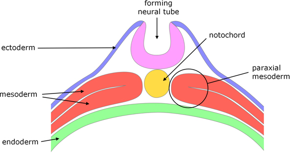
Figure 7. Formation of the neural tube by fusion of neural folds.
Closure begins approximately midway along the dorsal surface and progressively extends both posteriorly and anteriorly, but does not complete until after the first somites have formed (see below).
Neural crest cells
Whereas neural crest cells usually originate from the crests of the neural ridges before they meet (hence their name), in chondrichthyans they arise from the dorsal side of the neural tube after the ridges have fused and the neural tube has separated from the re-formed ectoderm.
Mesoderm
As mentioned above, at an early stage the mesoderm along the midline separates and forms the notochord, which then has a significant role in formation of the embryo. Immediately either side of the notochord the paraxial mesoderm (figure 7) differentiates into somites which form in pairs either side of the notochord, along the length of embryo, except for the head region. (Subsequently, the somites form the vertebrae.) Within the remaining ‘lateral plate’ mesoderm a cavity forms which effectively splits the mesoderm into two layers:
- inner ‘splanchnic mesoderm’ which overlies the endoderm (and subsequently forms the circulatory system), and
- outer ‘somatic mesoderm’ which lies within the ectoderm (and subsequently contributes to connective tissue).
Endoderm and formation of the gut
The edges of the embryonic shield – comprising the outer ectoderm, inner endoderm and both layers of the mesoderm – grow downwards (towards the yolk, in effect pushing the embryo upwards or away from the yolk), and then inwards so that the two sides move towards and then meet each other.

Figure 8. Formation of the primitive gut and main body cavity (coelom).
On meeting, each of the layers from each side fuses with the corresponding layer:
- first endoderm to form the primitive gut;
- then the mesoderm layers to produce the body cavity (around the gut) known as the coelom;
- and finally the ectoderm to complete the embryo’s external surface.
This takes place along the length of the embryo, except at the middle where the embryo connects with the yolk via an umbilicus which persists until the yolk is used up and the yolk sac is absorbed into the embryo.
Yolk membranes
Starting at about the same time as gastrulation, the margins of the blastula start to spread out (epiboly) across the yolk (the early stages of this can be seen in the ‘extraembryonic tissues’ identified in figure 6), gradually enveloping it, although this process is not complete until after considerable development has taken place (during Stage 4 in figure 9). All three germ layers overgrow the yolk such that the yolk sac is a trilamellar extension of the embryonic gut. However, the yolk membranes do not form a part of the developed embryo, except in so far as when the yolk is used up the membranes are absorbed into the embryo (unlike most cases where there are extraembryonic membranes which tend to be discarded).
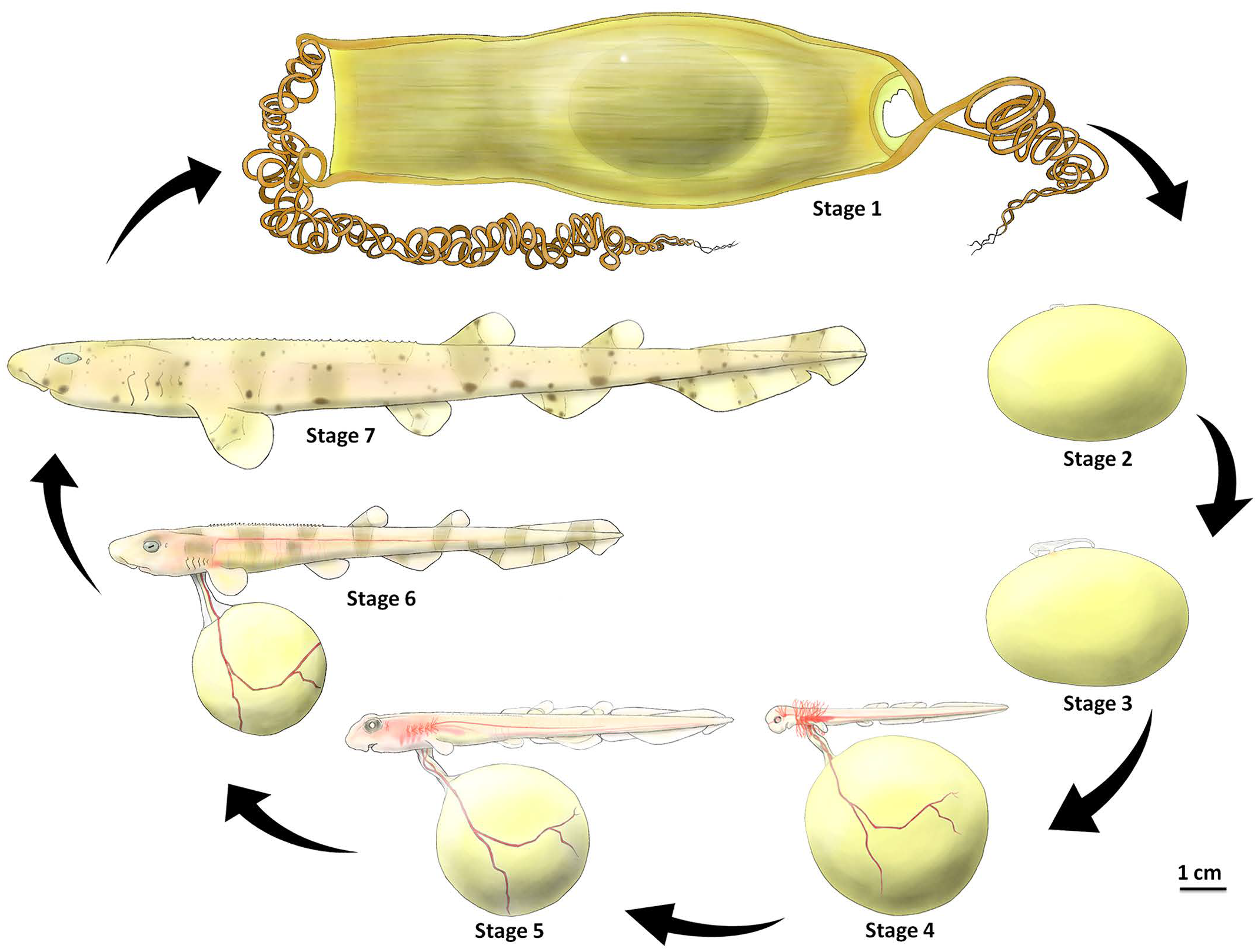
Figure 9. Overview of catshark embryonic development. [c]
Note that the stages assigned here do not correspond with the normal stages described by Ballard et al. [1].
Notes
1. William Ballard, Jean Mellinger and Henri Lechenaukt (1993), A Series of Normal Stages for Development of Scyliorhinus canicula, the Lesser Spotted Dogfish (Chondrichthyes: Scyliorhinidae), Journal of Experimental Zoology Vol. 267: 318-336.
Image credits
Graphics are by David Swift unless stated otherwise.
Background image for the page banner is by DrKontogianniIVF from www.needpix.com/photo/download/674083/embryo-ivf-icsi-infertility-fertility-free-pictures-free-photos-free-images-royalty-free.
a. Small-spotted catshark. Photo by Bj.schoenmakers. From https://commons.wikimedia.org/wiki/File:Scyliorhinus_canicula_(Small-spotted_catshark_or_dogfish),_Ecomare,_Texel,_the_Netherlands.JPG under the Creative Commons CC0 1.0 Universal Public Domain Dedication.
,_Ecomare,_Texel,_the_Netherlands.JPG)
b. Mermaids purse. Photo by Tom Oates, 2009. Image from https://commons.wikimedia.org/wiki/File:Mermaids_Purse.JPG under Creative Commons Attribution-Share Alike 3.0 Unported license.
c. Overview of catshark development. Figure 8 in Syafiq M. Musa, Molly V. Czachur, Holly A. Shiels (2018), Oviparous elasmobranch development inside the egg case in 7 key stages, PLoS ONE 13(11) e0206986 doi.org/10.1371/journal.pone.0206984. Open Access, distributed under the terms of the Creative Commons Attribution License.
Page created October 2020.