Comparison of the source tissues of embryonic germ layers in vertebrates
As mentioned in the comparison of gastrulation, a key outcome of gastrulation is the establishment of the germ layers – ectoderm, mesoderm and endoderm. The focus of this page is to point out that, as well as gastrulation being effected by different mechanisms, there are substantial differences in the source tissues of the germ layers. This is especially significant because the cardinal criterion for homology is that it should be the same structure, albeit in different forms. Recall from the introductory page on homology that two organs that appear the same but are derived from different embryonic sources are not homologous but analogous. So, although the germ layers in different vertebrate groups may appear equivalent, where they are derived from different embryonic sources they are not homologous.
In most cases, all three germ layers arise from an upper layer of cells of the blastula.
To avoid using ‘the germ-layer-forming layer’ or similar – on this page I refer to this layer as the ‘epiblast’, although this term is usually used only for the amniotes, and sometimes the teleosts.
However,
- the amphibian endoderm is a substantial exception to this;
- the areas of 'epiblast' becoming endoderm and mesoderm, or remaining as ectoderm, are the opposite way round between anamniotes and amniotes; and
- the significantly different ways in which it is formed (as outlined in the comparison of blastula formation) undermines any presumed homology of the 'epiblast' anyway.
Amphibian endoderm
The most striking exception to the general case that the germ layers arise from the 'epiblast' is that this is not true of amphibian endoderm. In amphibians, whereas the cells which become the ectoderm (the fate of remaining 'epiblast' cells after formation of the other germ layers) is the dome-shaped animal half of the blastula, the endoderm arises from the hemispherical vegetal half.

Figure 1. Fate map of Xenopus blastula, section.
Even in fish such as lamprey and bircher (not teleosts, see classification of the main groups of vertebrates), which have a blastula similar to amphibians, the endoderm arises from the peripheral or equatorial area of the 'epiblast', not the vegetal hemisphere. In these fish, the vegetal hemisphere produces extraembryonic tissues. [1]
Amniote versus anamniote sources of the germ layers
In anamniotes, gastrulation proceeds by involution or rolling over of cells at the edge of the blastoderm / 'epiblast'. The cells that involute or roll over then develop into endoderm (except for amphibians) and mesoderm. This means that the presumptive endoderm and mesoderm are around the periphery of the blastoderm / epiblast, and hence the cells that remain and become ectoderm are in the middle of the blastoderm.
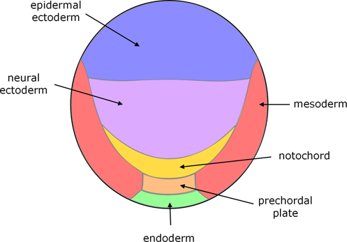
Figure 2. Fate map of catfish blastula; the blastula is substantially flat, and here is viewed from above.

Figure 3. Fate map of zebrafish blastula; the blastula is like a ball, and viewed here from the dorsal side, and somewhat from above.
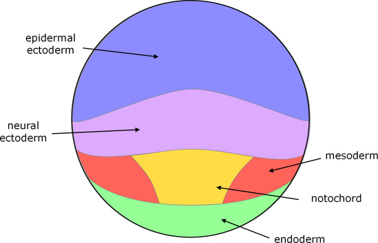
Figure 4. Fate map of Xenopus blastula; the blastula is like a ball, and viewed here from the dorsal side, and somewhat from above.
In contrast, in amniotes, the blastopore of reptiles and the primitive streak of birds and mammals is located in the middle of the epiblast. This means that the cells that become mesoderm and endoderm are derived from the middle area of the epiblast, and it is the peripheral cells which don’t involute or ingress that become ectoderm.

Figure 5. Fate map of turtle blastula, viewed from above.

Figure 6. Fate map of chick blastula, viewed from above.

Figure 7. Fate map of mouse blastocyst.
Hence, the fates of the superficial layer of the blastulas of anamniotes and amniotes are direct opposites. So this is a fundamental non-homology between amniotes and anamniotes.
Go to the Source tissues of germ layers in the Overview of diverse early embryonic development of vertebrates.
Notes
1. Masaki Takeuchi, Maiko Takahashi, Masataka Okabe, Shinichi Aizawa (2009); Germ layer patterning in bichir and lamprey; an insight into its evolution in vertebrates, Evolution of Developmental Control Mechanisms 332 p90-102.
Image credits
Graphics are by David Swift unless stated otherwise.
The background image for the page banner is taken from an image by ★Kumiko★ – https://www.flickr.com/photos/kmkmks/27388394090/, CC BY-SA 2.0, https://commons.wikimedia.org/w/index.php?curid=57660389
Page created October 2020.