Comparison of vertebrate blastulas
Embryos of different classes of vertebrates have very different blastulas, which of course arise in different ways, starting with different cleavage patterns which are discussed here.
Chondrichthyans
Following meroblastic cleavage, cell division continues to produce a mass of cells sitting within the cup of white yolk on top of the main body of yolk. These cells sort and differentiate to produce:
- an upper epithelial layer (surface blastomeres), which develops into the embryo,
- with the lower cells being mesenchymal, and
- at least some cells in contact with yolk form a yolk syncytial layer (YSL).
Beneath these is a cell-free zone – a subgerminal cavity – which becomes located at the posterior end of the embryo. (There is no blastocoel within the body of the cells.)

Figure 1. Catshark blastula.
Teleosts
Cleavage in teleosts is meroblastic and very stereotypical, and produces an array of cells positioned like a crown on top of the yolk. When there are about 128 cells, these sort and differentiate to produce three distinct cell populations:
- An outer enveloping layer (EVL) which is one cell thick. This has a significant role in formation of the embryo – it appears to provide the main driving force for epiboly – but does not become part of the embryo.
- The main component is a layer of deep cells. At first it is hemispherical or dome-shaped, but as epiboly proceeds this layer becomes of uniform thickness – initially about 4 cells thick, but reduces as it spreads during the course of epiboly and gastrulation. It is these deep cells that form the embryo.
- A yolk syncytial layer (YSL).
There is no blastocoel or subgerminal space.

Figure 2. Zebrafish blastula.
Amphibians
Cleavage in amphibians is holoblastic. A blastocoel begins during the first cell division, and after just a few divisions there is a clear segregation and differentiation of cells.
- The animal hemisphere has a dome of relatively small cells, comprising a one-cell-thick superficial layer overlying a deeper layer which is several cells thick. This dome of cells becomes the embryo's ectoderm and mesoderm.
- The vegetal hemisphere is occupied by relatively large cells which become the endoderm.
Note that the whole of the blastula becomes the embryo.

Figure 3. Xenopus blastula.
Reptiles
Following meroblastic cleavage the cells continue to proliferate to produce a mass of cells which sort and differentiate into:
- an upper epithelial epiblast, which is the main source of the embryo,
- a lower hypoblast, with a blastocoel in-between,
- marginal cells around the periphery, and
- a yolk syncytial layer (YSL).
There is a probably a subgerminal space below the hypoblast.
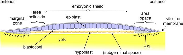
Figure 4. Turtle blastula.
Birds
Following meroblastic cleavage the cells continue to proliferate to produce a multi-layered disc of cells. These segregate and differentiate, and some cells are lost, to give:
- an upper single layer which becomes the epiblast, and subsequently is the main source of the embryo,
- beneath this are groups ('islands') of cells which precede the hypoblast (see below),
- marginal cells around the periphery, and
- a YSL, at least around the margins.

Figure 5. Early chick blastula.
- The hypoblast then grows from the posterior marginal cells, linking up the 'islands' of cells as it does so.

Figure 6. Chick blastula.
Mammals
In placental mammals, following rotational holoblastic cleavage, the cells continue to divide. At first the cells are loosely grouped within the zona pellucida, but when there are 8-16 cells they undergo compaction. Not only do they pack together more closely, but tight junctions form between them, a blastocoel begins to form, and the cells begin to differentiate. The blastocyst (mammalian blastula) comprises:
- outer trophoblast which develops into the placenta,
and an inner cell mass comprising -
- epiblast which becomes the embryo, and
- hypoblast which becomes extraembryonic tissues.

Figure 7. Human blastocyst.
The presumptive embryonic tissue, 'epiblast'
It will be apparent from the above outlines that there are significant differences between those parts of the blastula that will develop into the actual embryo. The term ‘epiblast’ is always used to describe this part in amniotes, quite often it is also called this in teleosts, but not usually in chondrichthyans or amphibians. But what it is called is much less important than the substance of what it is:
Chondrichthyans
It is a one-cell thick epithelial layer, forming the upper surface of the blastula.
Teleosts
It is a multiple-cell layer, not epithelial because it is beneath the EVL.
Amphibians
Unlike the other groups of vertebrates,
almost the whole of the blastula becomes the final embryo:
- the dome of the animal hemisphere, comprising a superficial single epithelial layer overlying a lower layer which is several cells thick, becomes the ectoderm;
- the mass of cells filling most of the vegetal hemisphere becomes the endoderm, and
- the cells at the margin of the animal dome and extending into the vegetal hemisphere, also comprising a superficial epithelial layer overlying deeper cells, become the mesoderm.
Reptiles and birds
It is a single-cell thick epithelial layer, overlying the hypoblast.
Mammals
It is part of the inner cell mass, within the outer trophoblast.
Hypoblast
The blastulas of amniotes have a further distinct layer of cells known as the hypoblast. Originally it was thought that this developed into the endoderm, but subsequently it was realised that it is substantially displaced (by involution in reptiles, ingression in birds and mammals) by cells from the epiblast, and it is mainly these that become endoderm. [1]
It should be noted that the hypoblast of amniotes:
- is part of the blastula,
- is distinct from the epiblast, and
- arises before gastrulation.
In anamniotes, it is common for superfical cells that are internalised in the course of gastrulation to be called hypoblast. But it should be noted that, in contrast to the hypoblast of amniotes, the ‘hypoblast’ of anamniotes:
- is derived from the superficial cells ('epiblast'),
- arises during gastrulation, and hence
- is part of the gastrula.
Go to Blastulas in the Overview of diverse early embryonic development of vertebrates.
Notes
1. More recent work has shown that, at least in birds, some cells of the hypoblast are not displaced, and become part of the definitive endoderm. Aaron Lawson, Gary Schoenwolf (2003); Epiblast and primitive-streak origins of the endoderm in the gastrulating chick embryo, Development 130 3491-3501.
Image credits
Graphics are by David Swift unless stated otherwise.
The background image for the page banner is taken from an image by ★Kumiko★ – https://www.flickr.com/photos/kmkmks/27388394090/, CC BY-SA 2.0, https://commons.wikimedia.org/w/index.php?curid=57660389
Page created October 2020.