Comparison of cleavage in vertebrates
The first divisions of the zygote are called cleavage. Cleavage is generally categorised as holoblastic, when the cell divisions pass right though the zygote or early embryo, or meroblastic when they do not. When meroblastic cleavage is limited to the germinal disc, it may be called discoidal.
Holoblastic cleavage
Cleavage is described as holoblastic where the first cell divisions pass right through the zygote or very early embryo. This is usually possible only where there is little yolk present.
Amphibians
In vertebrates, it is typified by amphibians because the first and second divisions are meridional, i.e. they extend through both of the embryonic poles, with the second being perpendicular to the first. The third cell division is perpendicular to both of the preceding ones; it is approximately equatorial but closer to the animal pole, reflecting increased yolk content towards the vegetal pole.

Figure 1. Initial cleavage in Xenopus.
Other features specific to amphibian cleavage are:
- the cortical rotation which precedes the first cell division, and
- the very early appearance of the blastocoel which begins to form between the two daughter cells during the first division.
Mammals (placental)
Holoblastic cleavage also occurs in placental mammals. What is unusual about cleavage in mammals is that, whereas the first division is meridional, the two second divisions are in different planes: one is meridional, and the other is equatorial. This is called rotational cleavage, and is unique among vertebrates.

Figure 2. Initial cleavage in placental mammals.
Meroblastic cleavage
Meroblastic cleavage is generally associated with the presence of substantial quantities of yolk. The cell divisions penetrate only through the germinal disc, they do not extend through the yolk. Usually, the first few cell divisions are perpendicular to the plane of the germinal disc, leading to a sheet of cells; and gradually divisions also occur parallel to the plane, leading to a clump of cells, as with chondrichthyans, or multiple layers of cells, as in birds and teleosts.
Chondrichthyans
In chondrichthyans, the first few divisions lead to a sheet of cells. When there are about 64 cells, further division gives rise to a clump of cells which begin to segregate and differentiate into an epithelial layer with mesenchymal cells beneath.
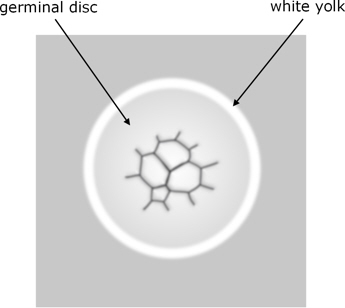
Figure 3. Early cleavage in chondrichthyans, viewed from above.
Birds
In birds, early cleavage produces a sheet of cells which is several cells thick.

Figure 4. Late cleavage in birds.
These then segregate (with loss of some cells) to produce the distinct cell populations of the blastula.
Reptiles
Early cleavage in reptiles is thought to be similar to that in birds, but subsequent segregation of the cells may be different.
Teleosts
In teleosts, the early cell divisions are very regular and stereotypical. The first five divisions are perpendicular to the plane of the germinal disc, with each division at right angles to the preceding one, leading to a regular array of 8 x 4 cells. The sixth division is then parallel to the plane of the germinal disc, bisecting all of the existing cells, to produce a 3-dimensional array of 2 x 8 x 4 cells. These appear rather like a crown on top of the yolk.

Figure 5. Stages of cleavage in teleosts. [a]
Evolutionary relationships of holoblastic and meroblastic cleavage
Mainly because cleavage in amphioxus (a fish-like invertebrate which is thought to resemble vertebrate ancestors) is holoblastic, this is generally reckoned to be the ancestral type of cleavage for vertebrates. It is presumed that meroblastic cleavage then evolved in response to increased quantities of yolk in the egg. Due to the distribution of meroblastic cleavage within the various vertebrate groups, it is inferred that this sort of transition occurred five times independently in different vertebrate lines (e.g. [1]). And further support for this multiple independent evolution is seen in the significant differences in the way meroblastic cleavage occurs, and in the subsequent blastula, in the different vertebrate groups.

Figure 6. Presumed phylogenetic distribution of holoblastic and meroblastic cleavage.
Symbol 0 indicates inferred evolution of meroblastic cleavage, X
where it reverted to holoblastic.
Hasley et al.[1] speculate on the benefits of meroblastic cleavage that could have driven its evolution and comment that, once evolved, it doesn’t revert to holoblastic. However, as with so many evolutionary inferences based on a cladistic or phylogenetic analysis, this ignores what would be have been required at the genetic and molecular levels in order to effect these changes – even once, never mind multiple times independently.
Accounts of how cleavage mechanisms are supposed to have evolved usually describe the general benefits of increased yolk in terms of the increased independence of the offspring. However, we should not forget that any increase in yolk and the associated structural changes to the egg would require genetic changes which are unlikely to occur anyway (in view of the formidable hurdles to evolving new genes). That is, it is an example of the challenge of genetics and molecular biology to any substantial evolutionary novelty.
Similarly, changing from holoblastic to meroblastic cleavage is usually depicted as a somewhat passive consequence of increased yolk, whereas in reality this too would have genetic implications. So, realistically, it is far more likely that the genetic and molecular implications that would be necessary to change from holoblastic to meroblastic cleavage, would preclude the other changes required to implement increased yolk content.
Also, it should be noted that cleavage in placental mammals is holoblastic, which implies that meroblastic would have had to revert to holoblastic in this case.
Go to Cleavage in the Overview of diverse early embryonic development of vertebrates.
Notes
1. Andrew Hasley, Shawn Chavez, Michael Danilchik, Martin Wühr, Francisco Pelegri (2017); Vertebrate Embryonic Cleavage Pattern Determination, chapter 4 in F. Pelegri et al. (eds.), Vertebrate Development, Advances in Experimental Medicine and Biology 953, DOI 10.1007/978-3-319-46095-6_4 , Springer International Publishing Switzerland.
Image credits
Graphics are by David Swift unless stated otherwise.
The background image for the page banner is taken from an image by ★Kumiko★ – https://www.flickr.com/photos/kmkmks/27388394090/, CC BY-SA 2.0, https://commons.wikimedia.org/w/index.php?curid=57660389
a. Cleavage in zebrafish. Author: Gilbert, SF. From https://en.m.wikipedia.org/wiki/File:Cleavage.png licensed under the Creative Commons Attribution-Share Alike 4.0 International license. Letters against each of the images have been blanked out.
Page created October 2020.