Comparison of somite formation in vertebrates
Somites are temporary embryonic structures which form from mesoderm. After they have formed, they differentiate into myotomal, sclerotomal and dermatomal tissues, which give rise to muscles, the axial skeleton including vertebrae, and to the dermis of the back, respectively. They occur on either side of the neural tube, the first pair forming at the anterior part of the trunk and the others forming sequentially from head to tail. The way this occurs is substantially the same in all vertebrates, except for amphibians for which it is significantly different.
Somitogenesis in most vertebrates
Following gastrulation, the notochord runs along the midline of the dorsal aspect of the embryo, and is flanked both sides by mesoderm. Soon after its formation the mesoderm on each side splits into two sheets or layers:
- an outer ‘somatic mesoderm’ which lies within the ectoderm, and
- an inner ‘splanchnic mesoderm’ which overlies the endoderm.
Close to the midline, sometimes these are referred to as myotome and dermatome because of their subsequent differentiation, e.g. as in Hamilton [1]. At first, and close to the notochord, these two layers are close together, but as they spread around the embryo, a substantial gap arises between them, which becomes the coelom. The mesoderm next to the notochord is called paraxial (or pre-somitic) mesoderm and it is this that transforms into the somites.

Figure 1. Section showing paraxial mesoderm, in birds.
The somites form by short lengths of the double layer of mesoderm pinching off, so the inner part is derived from splanchnic mesoderm (myotome) and the outer part from somatic mesoderm (dermotome), and the gap in between gives rise to a space in the centre of the somite called the myocoel (see below). The mesoderm is mesenchymal, but the surface of the somites is epithelial, so the morphological formation of somites involves a mesenchymal-to-epithelial transition.
Somite formation in amphibians
In amphibians, following gastrulation the relationship of the ectoderm, notochord and mesoderm is similar to that of tetrapods (whilst noting the bi-layered ectoderm).

Figure 2. Section showing paraxial mesoderm in Xenopus.
However, not only does the neural tube form very differently, but the somites form from only the inner (splanchnic) mesoderm. Consequently, there is no myocoel, and the outer (somatic) mesoderm remains as a separate layer on the outside of all of the somites.
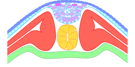
Figure 3. Secton showing formation of somites from inner (splanchnic) mesoderm in Xenopus.
Further, the somites do not ‘pinch off’ from the mesoderm (as in most vertebrates) but form by cells of the inner layer rotating though 90° so that they become orientated parallel to the neural tube / notochord – whereas in other vertebrates the cells remained aligned perpendicular to it. These significant differences are indicated in Figure 4 which is a composite – comparing what happens in an amphibian (Xenopus laevis) on the left with the usual vertebrate /tetrapod on the right.

Figure 4. Comparison of somite formation in amphibian (Xenopus laevis) and typical tetrapod. Longitudinal section (a) showing progressive formation of somites, and cross-section (b) through newly-formed somites. [a]
As well as the differences depicted here, amphibian somites are not epithelial, so there is no mesenchymal-to-epithelial transition.
Hamilton sums up the situation as follows:
The presumptive somitic material of the early neurula and the fully differentiated axial musculature and dermis of Xenopus appear to be very similar to those of other vertebrates [which is why they are generally considered to be homologous]. Yet the intervening stages of morphogenesis are strikingly different [which refutes that presumed homology]. [1]
Go to Somitogenesis in the Overview of diverse early embryonic development of vertebrates.
Notes
1. Louie Hamilton (1969); The formation of somites in Xenopus, J. Embryol. exp. Morph 22(2) pp253-64.
Image credits
Graphics are by David Swift unless stated otherwise.
The background image for the page banner is taken from an image by ★Kumiko★ – https://www.flickr.com/photos/kmkmks/27388394090/, CC BY-SA 2.0, https://commons.wikimedia.org/w/index.php?curid=57660389
a. Figure 4 is based on figure 5 in Hamilton [1].
Page created October 2020.