Comparison of neural tube formation in vertebrates
Terminology: 'primary' and 'secondary' neurulation — and why I don’t use these!
- In amniotes and some other vertebrates, the main part of the neural tube arises by formation and fusion of folds of neural ectoderm, a process which is sometimes called primary neurulation. In this case, the underlying notochord forms first, and is thought to have a significant role in the development of the neural tube.
- In at least birds and mammals (and probably amphibians), the sacral region of the neural tube forms very differently. It develops from mesoderm (not ectoderm), from cells which are initially mesenchymal in nature, but then condense to form a rod of tissue (the medullary cord). The lumen of the neural tube then arises within this rod by cavitation, extending the ectoderm-derived neural tube. This process is often referred to as secondary neurulation. In this case, the notochord develops in parallel with, may even lag behind, the overlying neural tube.
In teleosts (most bony fish) the neural tube forms from neural ectoderm, but which first thickens, and then the lumen of the neural tube arises by cavitation within that thickening; and the notochord forms before the neural tube. Because it forms by cavitation, this process is also sometimes referred to as secondary neurulation; but it is clearly very different from the ‘secondary neurulation’ mentioned above.
Further, in amphibians, although the main part of the neural tube forms from ectoderm, it does so by a process which partly resembles the coming together of ectoderm folds (and the notochord forms before the neural tube), but also by cavitation of thickened ectoderm. It is clearly different from either the ‘primary’ or ‘secondary’ neurulation described above, and there seems little point in debating whether it is one or the other.
So to avoid confusion or being misleading, in the following I do not use the terms ‘primary’ or ‘secondary’ neurulation.
And I consider development of only the main part of the neural tube, not the sacral part.
Fusion of ectoderm folds
In most vertebrate groups (amniotes and cartilaginous fish), the neural tube arises by the formation and fusion of folds of the neural ectoderm. The neural ectoderm comprises a single layer of cells which is epithelial in nature, and which thickens and narrows – convergent extension – and by its cells changing shape from cuboidal to columnar. The edges of the neural plate then rise to form neural ridges which meet and fuse on the dorsal side of the embryo, enclosing the neural tube.

Figure 1. Section to show mechanism of neurulation in birds.
Folding
The neural ridges fold by means of specific hinge points which run along the length of the neural plate. The cells at these hinge points promote bending of the neural epithelium because they become triangular in cross-section – narrower on the side that will become the lumen of the neural tube. These cells can be identified from about the same time that the neural ridges begin to rise.
Some of the force driving closure of the tube is also thought to be from expansion of the ectoderm either side of the neural ridges.
Fusion
It would be easy to think that fusion of the folds would be straightforward. However, fusion is mediated by specific molecules being produced on the cells at the tips of the folds, which interact and bring the two folds together. It is also important to note that the folds need to be aligned correctly so that the non-neural ectoderm on either side, and the lining of the neural tube on either side, are united correctly.
Clearly, with this process of neurulation, formation of the neural tube and closure of the final epidermis are necessarily linked and simultaneous.
Fusion generally starts at 1, 2 or 3 specific points along the embryo’s axis, and then proceeds (zipper-like) in both directions from each of these initial points.
Epithelium
It should be noted that the dorsal surface of the neural plate, including what will become the inner surface of the neural tube, is a polarised epithelium, and remains epithelial in nature throughout the process of neurulation, except transiently at the cells that implement fusion of the folds.
Neural crest cells
Progenitors of the neural crest cells are located at the edges of the neural plate, at its boundary with non-neural ectoderm. So when the neural folds meet they are positioned at the crests of the folds – which gives these cells their name. Usually these cells become incorporated into the dorsal surface of the neural tube before migrating elsewhere.
Cavitation within a thickening of the ectoderm
In teleosts (the most abundant group of fish) the neural tube forms very differently.
The single-layered neural plate converges towards the midline, briefly forming a double layer of superficial and deep cells. The deep cells fold inwards along the midline, and the superficial cells intercalate with the deep ones, to form a wedge-shaped mass of cells called the neural keel.

Figure 3. Zebrafish neural keel
The keel then adopts a cylindrical or rod-like cross-section before separating from the ectoderm which reforms behind it, and becomes epidermis. Only after this rod has separated from the epidermis does a lumen form – by cavitation – within the rod.
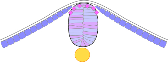
Figure 4. Zebrafish neural rod. [same colour on opposite sides?]
The cells within the neural rod become rearranged into right and left groups facing the midline. A notable feature of lumen formation is that at the rod stage cells on either/each side of the midline divide, with the daughter cells separating either side of the midline, i.e. one cell ending up on one side of the lumen, and the other on the opposite side – called mirror-symmetric cell division. Almost all of the cells of the early neural tube arise in this way. As a result, both sides of the neural tube receive equal contributions from both left and right sides of the neural plate. The initial cavity is a slot which arises between the left and right halves, perpendicular to the dorsal ectoderm, and this slot then widens to form the lumen of the neural tube.
Neural crest cells
When the neural tube forms this way it does not involve neural folds, so there are no neural crests. Nevertheless, characteristic ‘neural crest cells’ arise from the lateral border of the neural plate, migrate through the neural keel and become part of the dorsal neural tube, before migrating elsewhere.
Comparison with neurulation by the fusion of ectoderm folds
Neurulation by cavitation in teleosts differs from that by the fusion of ectoderm folds in the following ways:
- Most obviously, there is no formation of folds or ridges of ectoderm;
- so there is no fusion of folds.
- Whereas in amniotes the formation of the neural tube and epidermis are linked, in teleosts these processes are separated and independent.
- In amniotes the epithelial nature of the ectoderm is retained through to the lumen of the neural tube; whereas in teleosts, a polarised epithelium must be formed within the neural keel, by mesenchymal-to-epithelial transition.
- In amniotes each side of the neural tube is derived from the respective side of the neural plate; whereas in teleosts both sides of the neural tube are derived more-or-less equally from both sides of the neural plate.
Neurulation in amphibians
In amphibians the neural tube forms in a way which resembles yet differs from both fusion of ectoderm folds and cavitation within a keel of ectoderm cells.
Unlike the other vertebrate groups mentioned, from the outset the amphibian neural plate comprises two layers: an upper epithelial layer and a lower mesenchymal layer, and these reform towards the end of neurulation. In the course of neurulation both layers of cells converge towards the midline to produce a neural groove, flanked by shallow ridges, on the surface, and bulge or keel of cells between that and the notochord.

Figure 4. Xenopus neural groove stage.
The sides of the groove fuse, almost simultaneously along the full length of the neural groove, usually enclosing a small lumen, although its surrounding cells are mesenchymal rather than epithelial. However, they gradually organise into a clearly defined neural tube, involving a change from mesenchymal to epithelial cells. And as the neural tube forms, a deep layer of dorsal epidermis reforms over the neural tube (and below the superficial epidermis).
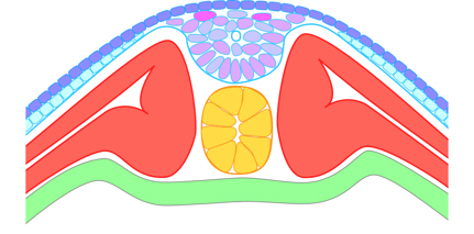
Figure 4. Xenopus after closure of the neural groove.
Comparison with amniote fusion and teleost cavitation
Neurulation in amphibians resembles that in amniotes in so far as it involves fusion of neural ridges either side of a groove and usually (but not always) encloses a small lumen; and it resembles teleost cavitation within a thickening of the ectoderm-derived cells. However, there are marked differences from both:
Differences from fusion
- Whereas in amniotes the ectoderm is a single epithelial layer of cells, in amphibians there is also a lower mesenchymal layer, and both layers reform after formation of the neural tube.
- Whereas in amniotes fusion of the neural folds starts at discrete points and then propagates from these, in amphibians the sides of the neural groove fuse almost simultaneously along its entire length.
- Whereas, in amniotes, fusion always results in formation of a lumen – because fusion is how the neural tube is formed – this is not always the case in amphibians.
- Whereas in amniotes the internal surface of the neural tube lumen is epithelial right from the outset, in amphibians the cells surrounding the early lumen (if present) are mesenchymal.
- Similarly, whereas in amniotes fusion results immediately in a formed neural tube, in amphibians the cells below the point of fusion are disorganised, and the definitive lumen is reconstructed from these over a period of several hours.
Differences from cavitation in teleosts
- Whereas in teleosts the ectoderm is a single epithelial layer of cells, in amphibians there is also a lower mesenchymal layer. Both layers reform after formation of the neural tube.
- In teleosts there are no ectodermal fold or ridges, so no fusion of these.
- In amphibians fusion usually results in the formation of a small lumen, which does not occur in the neural keel of teleosts.
- In amphibians, there is no mirror-symmetric cell division which is characteristic of teleost neurulation.
Go to Neurulation in the Overview of diverse early embryonic development of vertebrates.
Notes
Notes display in the main text when the cursor is on the Note number.
Image credits
Graphics are by David Swift unless stated otherwise.
The background image for the page banner is taken from an image by ★Kumiko★ – https://www.flickr.com/photos/kmkmks/27388394090/, CC BY-SA 2.0, https://commons.wikimedia.org/w/index.php?curid=57660389
Page created October 2020.