Comparison of the formation of vertebrate extraembryonic membranes
As the term implies, extraembryonic membranes refers to various membranes that form in the course of embryonic development but do not form part of the final embryo. Usually this means the yolk sac, amnion, chorion and allantois of the amniotes – reptiles, birds and mammals.
Anamniote yolk sac
Anamniotes with a yolk – notably the cartilaginous and teleost fish – also have a yolk sac which grows over the yolk. In teleosts this arises as part of gastrulation and is part of the embryo, so it is not extraembryonic.
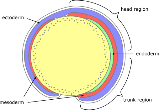
Figure 1. Zebrafish yolk sac.
In chondichthyians the yolk sac grows over the yolk after gastrulation. Although the yolk sac is outside the body of the embryo, as the yolk is used up the sac diminishes, and eventually the yolk sac is reabsorbed into the maturing embryo, which does not occur with the extraembryonic membranes of amniotes.
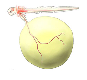
Figure 2. Catshark yolk sac.
Amniote extraembryonic membranes
The extraembryonic membranes of amniotes form in two very different ways:
Fusion of folds that arise after gastrulation
In almost all amniotes, including most mammals, the extraembryonic membranes arise after gastrulation as folds of tissue which arise around the periphery of the embryo and fuse above (dorsal) and below (ventral) the embryo.
Birds
For example, in birds, the dorsal folds include ectoderm and somatic mesoderm. When these folds meet above the embryo they form:
- an inner amniotic cavity which surrounds the embryo; and
- an outer chorionic cavity which is lined with extraembryonic mesoderm and in effect is an extension of the embryonic coelom (and sometimes is called the extraembryonic coelom).
Note that, although both of these membranes comprise layers of ectoderm and mesoderm, the order of these layers in relation to the cavity they enclose is reversed.

Figure 3. Chick extraembryonic membranes.
The ventral folds comprise ectoderm and somatic mesoderm on their inner surface, with endoderm and splanchnic mesoderm on their outer surface. When these meet below the embryo:
- the ectoderm and mesoderm complete formation of the chorionic cavity; and
- the endoderm and mesoderm surround the yolk, to form the yolk sac.
A further extension of the endoderm and mesoderm, from the hind-gut region, forms an additional cavity called the allantois which collects waste products from the embryo. As the embryo develops, the yolk shrinks and the allantois grows until it completely surrounds the chorionic cavity.
Mammals
In most mammals, the extraembryonic membranes form in a similar way to reptiles and birds, but with some differences because of the structure of the mammalian blastocyst, which includes a trophoblast, and the formation of a placenta.
For example, in rabbit, after gastrulation, folds of ectoderm and mesoderm arise laterally and posteriorly (but little if any anteriorly); and when these folds meet dorsally, they form the amnion and its cavity, and part of the chorion.

Figure 3. Rabbit extraembryonic membranes.
Ventrally,
- the cavity within the trophoblast, which was part of the blastocyst, persists through gastrulation and becomes the yolk sac, with the trophoblast becoming coextensive with the ectoderm;
- the endoderm of the extraembryonic folds spreads around the inside of the yolk sac, but this is not complete until the allantois and placenta have formed.
As in birds, a pocket of endoderm and mesoderm in the hindgut regions forms the allantois.
In most mammals the membrane of the allantois and the chorion combine to form a placenta.
Cavitation of parts of the blastocyst (before gastrulation)
Primates
In humans, and it is thought to be the case in all primates, formation of the extraembryonic membranes is radically different:
- all of the extraembryonic cavities arise by cavitation rather than by the fusion of folds;
- they all form before gastrulation and
- there is a key role for the pre-gastrulation extraembryonic mesoderm which is absent in reptiles and birds.
1. The first cavity to arise is the the blastocoel of the blastocyst, which becomes the (primitive) yolk sac, lined with cells which spread from the hypoblast. (This resembles the spread of endoderm in mammals such as the rabbit; but whereas this does not occur in the rabbit until after gastrulation and is not complete until much later, in primates even the secondary yolk sac is formed before gastrulation begins.)
2. The amnion arises through cavitation of the epiblast.
3. Extraembryonic mesoderm is a layer of tissue that arises between the lining of the primary yolk sac and the cytotrophoblast, and spreads to cover the amniotic cavity as well. As this tissue thickens, cavities form within it, and coalesce to form the chorionic cavity which consequently is lined with extraembryonic mesoderm.
4. Some of this extraembryonic mesoderm also forms the outer layer of the secondary yolk sac.
5. And the allantois arises as an outpocketing of the secondary yolk sac, but is generally small in primates.

Figure 3. Human extraembryonic membranes.
All of this occurs before gastrulation, and there is no formation or fusion of folds of germ layers.
Conclusion
This is one of the most striking anomalies from an evolutionary perspective:
- The morphological differences between these two developmental mechanisms are so substantial;
- to change from the usual fusion of post-gastrulation folds to cavitation within the pre-gastrulation blastula, would entail changes at such an early stage of development; and
- would need to have occurred within such a recently evolved group as the placental mammals.
Go to Extraembryonic membranes in the Overview of diverse early embryonic development of vertebrates.
Notes
Notes display in the main text when the cursor is on the Note number.
Image credits
Graphics are by David Swift unless stated otherwise.
The background image for the page banner is taken from an image by ★Kumiko★ – https://www.flickr.com/photos/kmkmks/27388394090/, CC BY-SA 2.0, https://commons.wikimedia.org/w/index.php?curid=57660389
Page created October 2020.