Embryonic development of mammals (humans)
Humans are classified within the group of mammals called eutherians which (with a few exceptions) use a placenta to nourish the developing embryo within the mother.
The other main groups of present-day mammals are:
- marsupials (pouched), and
- monotremes (egg-laying).
Egg and fertilisation
The human ovum comprises a single cell, about 0.1 mm in size, containing the haploid nucleus. It is enveloped by a clear jelly-like coat called the zona pellucida; and the whole is surrounded by a population of follicular cells (originating from the ovary) which form the corona radiata.

Figure 1. Section through human ovum.
Fertilisation takes place in the upper region of the oviduct (Fallopian tube) and the early stages of development unfold as the embryo travels along the oviduct. During its passage along the oviduct, the embryo loses some of the cells of the corona radiata, but the zona pelludica remains intact. Implantation in the uterus occurs when development has reached the blastocyst (blastula) stage.
Cleavage to blastocyst
Consistent with the absence of a significant yolk, cleavage in mammals is holoblastic, meaning that the first division extends right through the egg cell. The first division is typical of other organisms having holoblastic cleavage, with the division being meridional – extending from one pole to the other. However, in mammals the second cleavage is unusual: one cell divides meridionally, but the other divides equatorially; which is called rotational cleavage.

Figure 2. Rotational cleavage: cleavage 2a is meridonal, 2b is equatorial.
In most other organisms (having holoblastic cleavage) both of the second divisions are meridional, and the third or subsequent divisions are equatorial.
Also, whereas in other vertebrates the early cell divisions tend to be synchronous, this is often not the case with mammals, such that there is an odd number of cells instead of the usual geometric increase 2, 4, 8 … .
Early cell divisions also tend to be slower than for other animals.
The cell divisions up to just before implantation take place within the zona pellucida, so there is no overall increase in size of the embryo but its cells become smaller as they proliferate.
Compaction and morula
Up to 8 – 16 cells, they form a loose association within the zona pellucida, but then they compact, with tight junctions forming between the outer cells. This is called the morula stage.
It is about now that the embryonic genome is activated, and this is followed by the first clear differentiation of cells, with further cell divisions resulting in inner cells being distinct from the peripheral ones, and the beginning of a fluid-filled cavity, the blastocoel.
Blastocyst
The blastocyst is the mammalian equivalent of the blastula in other vertebrates. It comprises three populations of cells:
- an outer cell layer (trophoblast) which develops into the placenta; and
an inner cell mass:
- most of which is the epiblast, which is the source of embryonic tissues, and the amnion,
- although the layer of cells in contact with the blastocoel is the hypoblast, most of which forms extraembryonic tissues.

Figure 3. Human blastocyst.
Implantation and embryonic disc
About 7 days after fertilisation the embryo loses the zona pellucida, and implants into the lining of the uterus, by which time it comprises about 200 cells.
Around the time of implantation:
- the amniotic cavity arises within the epiblast,
- the blastocyst cavity becomes the primary yolk sac, which is lined by cells that spread from the hypoblast.
In addition, cells from the epiblast and hypoblast organise into a two-layered structure known as the embryonic disc, positioned between the primary yolk sac and amniotic cavity. Previously these two layers had been thought to correspond with the first two germ layers, the ectoderm and endoderm, but see below.
The trophoblast penetrates the uterine wall and begins to form the placenta; at an early stage it differentiates into two distinct layers, known as the cytotrophoblast and the syncytiotrophoblast.

Figure 4. Embryo shortly after implantation, with embryonic disc.
Extraembryonic mesoderm
Extraembryonic mesoderm is a layer of tissue that arises between the lining of the primary yolk sac and the cytotrophoblast, and spreads to cover the amniotic cavity as well. As this tissue thickens, cavities form within it, and coalesce to form the chorionic cavity which is lined with extraembryonic mesoderm (figure 5). (Note that this is different from the germ-layer mesoderm which forms later, see below.) In this process some of the primary yolk sac is lost, and what remains is called the secondary yolk sac. The embryo remains attached to the internal lining of the chorion by a connecting stalk of extraembryonic mesoderm which becomes the umbilical cord.

Figure 5. Longitudinal section pre-gastrulation (approx. day 14).
Gastrulation
From about day 14 after fertilisation, a groove appears near the caudal end of the epiblast, it extends about two-thirds of the way along the midline towards the cranial end, terminating in a widening with a depression at its centre. This primitive groove and primitive pit are where gastrulation occurs. As gastrulation proceeds, the primitive node recedes caudally, with a corresponding shortening of the primitive streak.
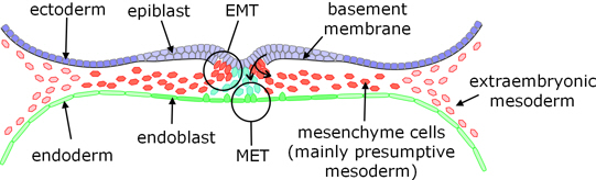
Figure 6. Transverse section (perpendicular to figure 5) through the embryonic disc, showing gastrulation.
In the course of gastrulation, cells of the epiblast proliferate and move towards the groove where they transition from epithelial to mesenchymal in character, and ingress below the surface. In the early phase, these ingressing cells enter the hypoblast, reverting to epithelial cells (mesenchymal to epithelial transition) to become the definitive endoderm; at the same time displacing the hypoblast cells from the embryonic disc to line the yolk sac. As this stage proceeds, further cells ingressing from the epiblast move into the space between the epiblast and endoderm to form a middle layer of cells called mesoderm. (At the edges of the embryonic disc this embryonic mesoderm merges with the previously formed extraembryonic mesoderm.) Once the mesoderm is formed, the remaining epiblast is called ectoderm, and the three germ layers are complete.
Notochord
The notochord forms within the mesoderm in three stages [1]:
- First, the primitive pit extends into the mesoderm, below the epiblast / ectoderm, towards the cranial end of the embryonic disc, resulting in a hollow tube called the notochordal process (or canal).
- This process merges with the underlying endoderm, to form the notochordal plate (during the early stages of this process, there is therefore a connection from the overlying amniotic cavity through to the underlying yolk sac via the lumen of the notochordal process, this connection being called the neuroenteric canal although in other contexts this term generally refers to a connection via the forming neural tube and the forming gut).
- Then, after about a day, the definitive notochord (which is a solid rod, rather than a tube) emerges from the notochordal plate.
Throughout this development, cells of the presumptive or definitive notochord are in contact with the overlying epiblast / ectoderm, and stimulate it to develop the neural tube.

Figure 7. Transverse section following gastrulation, approximately through the middle of the embryonic disc, showing prenotochordal process /canal, and the beginning of the neural ridges.
Neural tube
An early role for the notochord, including its precursors, is to induce formation of the neural tube. As gastrulation completes, ectoderm converges towards and thickens along the midline to form the neural plate. The lateral edges of the neural plate rise to form ridges (with the neural groove in-between) which meet and fuse to enclose the neural tube.
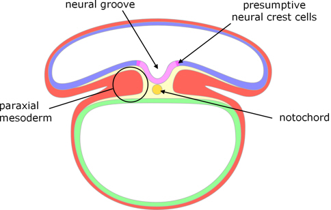
Figure 8. Transverse section showing formation of the neural tube.
Neural crest cells
Cells of the ectoderm that line the edges of the neural groove are called neural crest cells. When the neural tube closes these cells become separated from both the ectoderm and the neural tube, initially located between them, but subsequently migrate to form various tissues.
Mesoderm
Either side of the notochord the (embryonic) mesoderm splits into a double layer:
- the ‘somatic mesoderm’ which underlies the ectoderm (and subsequently contributes to connective tissue), and
- the ‘splanchnic mesoderm’ which overlies the endoderm (and subsequently forms the circulatory system).
At the edges of the embryonic disc, these layers of embryonic mesoderm merge respectively with:
- the extraembryonic mesoderm of the amnion; and
- the extraembryonic mesoderm that lines the yolk sac.
Somites
Immediately either side of the notochord, what is known as the paraxial mesoderm segments into somites which form as pairs either side of the notochord.

Figure 9. Transverse section showing formation of the gut.
Coelom and gut
While the neural tube and somites are forming, the amnion begins to spread ventrally around the embryo. When the folds of membranes meet and fuse:
- they pinch off part of the yolk sac, which becomes the primitive gut, having an internal layer of endoderm;
- the spaces between the two layers of mesoderm coalesce to form the coelom or body cavity; and
- the embryo is enveloped by the amniotic cavity.
Extraembryonic membranes
It will be apparent from the foregoing description that the extraembryonic membranes, and the cavities they enclose, arise at an early stage in embryonic development: notably, before gastrulation.
Notes
1. Karel de Bree, Bernadette S. de Bakker, Roelof-Jan Oostra (2018); The development of the human notochord, PLoS ONE 13(10): e0205752, doi: 10.1371/journal.pone.0205752
Image credits
Graphics are by David Swift unless stated otherwise.
Background image for the page banner is by DrKontogianniIVF from www.needpix.com/photo/download/674083/embryo-ivf-icsi-infertility-fertility-free-pictures-free-photos-free-images-royalty-free.
Page created October 2020.